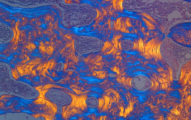
The Hidden Beauty of Disease Under Our Skin
Beneath our sheath of skin is an internal world both vast and complex. While most of us rarely get to see it, these workings of our systems and organs are the daily viewing of pathologists, particularly when it comes to disease. A new book of photography takes us into our own interiors, and shows th

Beneath our sheath of skin is an internal world both vast and complex. While most of us rarely get to see it, these workings of our systems and organs are the daily viewing of pathologists, particularly when it comes to disease. A new book of photography takes us into our own interiors, and shows that even with their horrid ravaging of our bodies, there is some beauty in these afflictions.
“Let me be very clear, in no way do we mean to glorify disease, but it is part of the human condition,” co-author of Hidden Beauty: Exploring the Aesthetics of Medical Science Norman Barker explained. Yet he elaborated that we’ve all experienced diseases in some way, whether in our own lives or someone we know. Cancer, Alzheimer’s, kidney disease, heart attacks, and infections are not unfamiliar to us, but their structure in our bodies is often invisible.
Barker, who is Associate Professor of Pathology and Art as Applied to Medicine at the Johns Hopkins University, collaborated on the book with Christine Iacobuzio-Donahue, a professor of pathology, oncology, and surgery also at Johns Hopkins. His job title might seem curious, but he noted that art and anatomy “have always had a close relationship” and that “during the Renaissance artists and anatomists were often the same person.” For example, he pointed out that one of the most famous drawings of the 16th century was the frontispiece of anatomist Andreas Vesalius’s De Humani Corporis Fabrica (1543), showing the dissection by Vesalius of a woman’s corpse. Later, people would pay to enter Dutch anatomy theaters to see the interior of human bodies. This interest hasn’t left us, even if the cross between direct research on the human body and art is not as close as it once was, and science remains a very visual study.
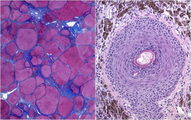
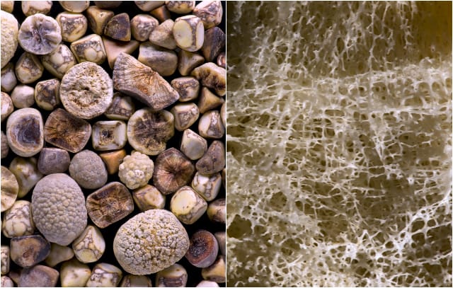
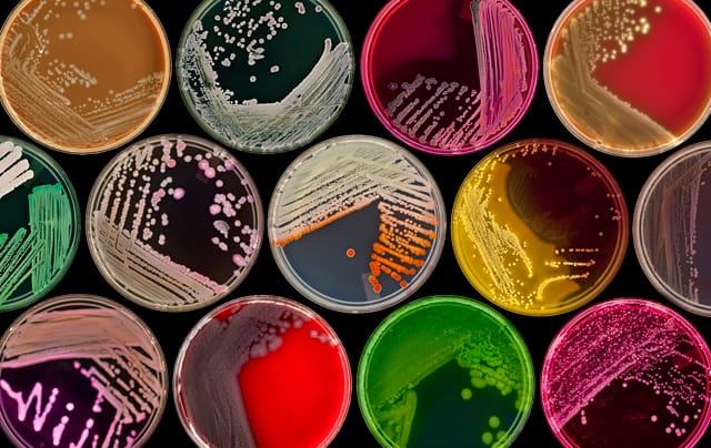
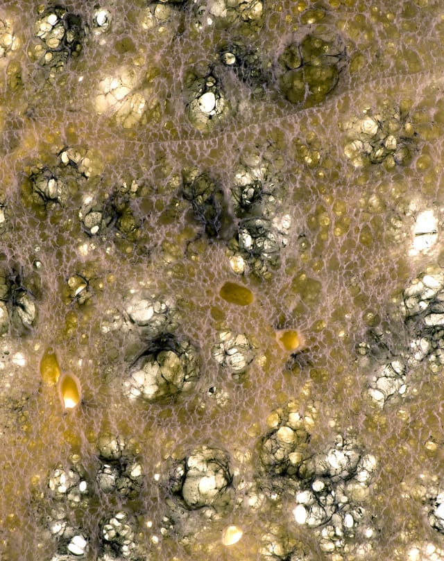
Photographs from Hidden Beauty are currently being exhibited the Johns Hopkins University School of Medicine and will later travel to the Mütter Museum in Philadelphia. They include a variety of imaging techniques, such as a cancer cells revealed by Spectral Karyotyping or electron microscopy. While the original intention wasn’t that they be visual art, the diagnosis images have striking views of forms and a diversity of structures, so that it’s astounding that they all came from the human body. Nevertheless, the goal of the book is not just to show the entrancing dimensions of such things as a lung riddled by smoke inhalation or the worn lattices of osteoporosis, but to explain the diseases in an accessible context as well. It’s by really seeing these diseases and the inner-workings of ourselves that we can have a better understanding of what’s happening beneath our surfaces.
“The human body is a magnificently complicated machine,” Barker stated. “We feel privileged to have a behind the scene tour of the human body everyday. We also feel privileged to work with men and women who have devoted their lives to science and find a cure for many of these diseases.”
Hidden Beauty: Exploring the Aesthetics of Medical Science is available from Schiffer Publishing.
