This Close-Up of an Ant’s Face Will Scar You Forever
The image is one of many in the Nikon Small World Photomicrography Competition that reveals the devil is definitely in the details.

What you see before you is not, as you might think, someone's nightmarish Halloween imaginings, but an actual picture of an actual ant captured at 5X macro range. Despite what Pixar would have us believe, ants are apparently hellish gremlins more at home in the Alien franchise than A Bug's Life. This is just one of many revelations delivered by the 48th Annual Nikon Small World Photomicrography Competition, an event that reveals the devil is definitely in the details.
https://www.instagram.com/p/CNUKYnFgkdZ/
This year's top award went to Grigorii Timin and Dr. Michel Milinkovitch of the University of Geneva, who submitted a micro-glimpse into the embryonic hand of a Madagascar giant day gecko. The image shows the complex interior of the gecko hand with some fingers jointed in three or four places, giving the creature the incredible dexterity it needs to navigate tree branches with ease. The ribbed outline of the fingers and the inner vasculature are all visible in the photo, which was generated using confocal microscopy at an incredible 63X range.
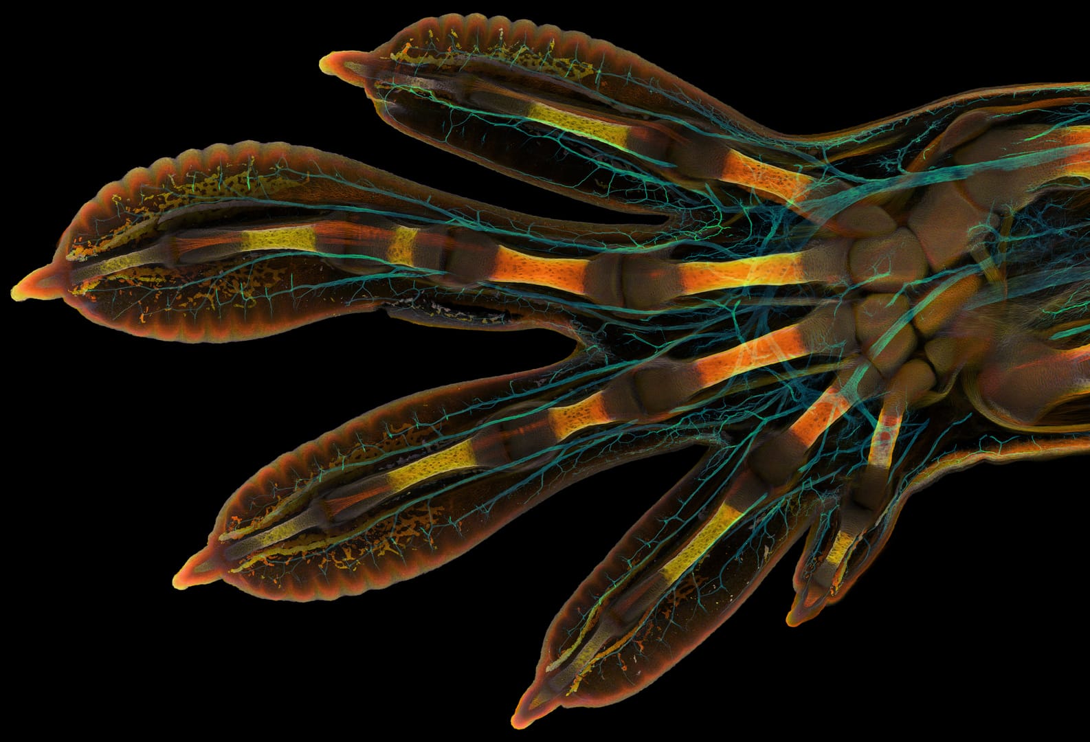
“This embryonic hand is about 3 mm (0.12 in) in length, which is a huge sample for high-resolution microscopy,” Timin said in a statement. “The scan consists of 300 tiles, each containing about 250 optical sections, resulting in more than two days of acquisition and approximately 200 GB of data.”
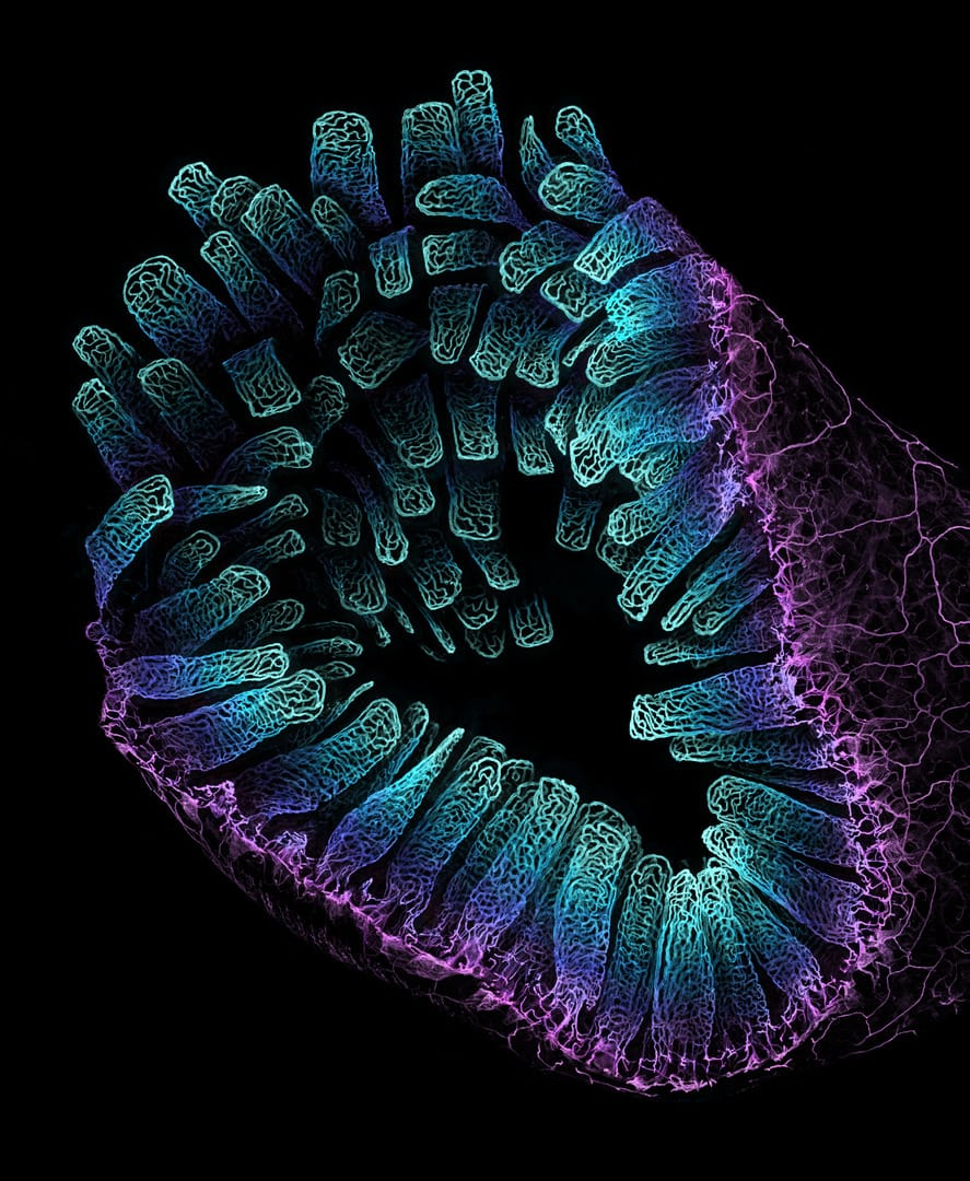
If you're anything like me, you constantly wonder what the blood vessel networks look like in the intestine of an adult mouse. Wonder no more! Third place was awarded to Satu Paavonsalo and Sinem Karaman from the University of Helsinki, for their cool-hued, anemone-like micro-image of just such a subject.
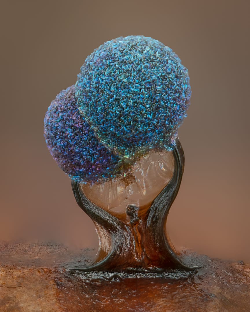
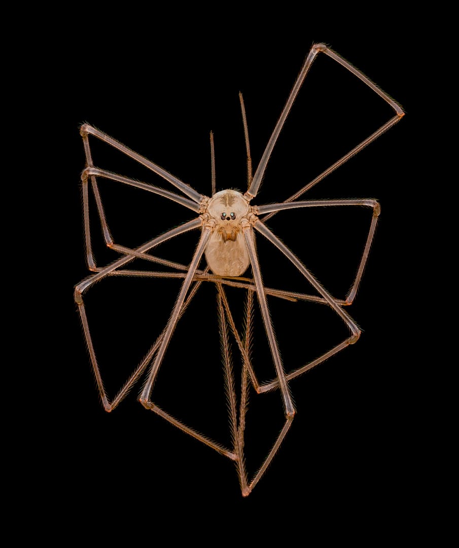
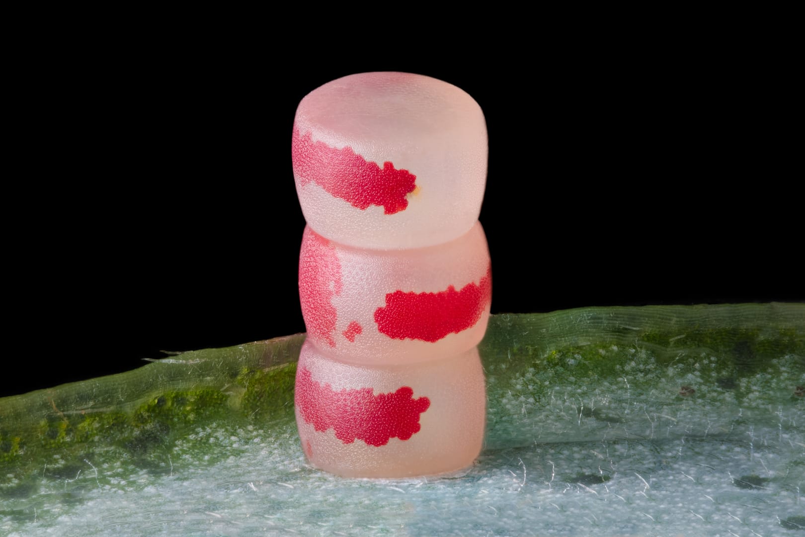
From a stack of moth eggs that look like a perfect minimalist sculpture and a close encounter with a daddy long-legs spider (which is, for the record, still an order of magnitude less terrifying than that ant) to a presentation of slime mold that would be perfectly at home decorating a Christian ministry with new-age vibes, these incredible images show the true artistry of nature at its most detailed.
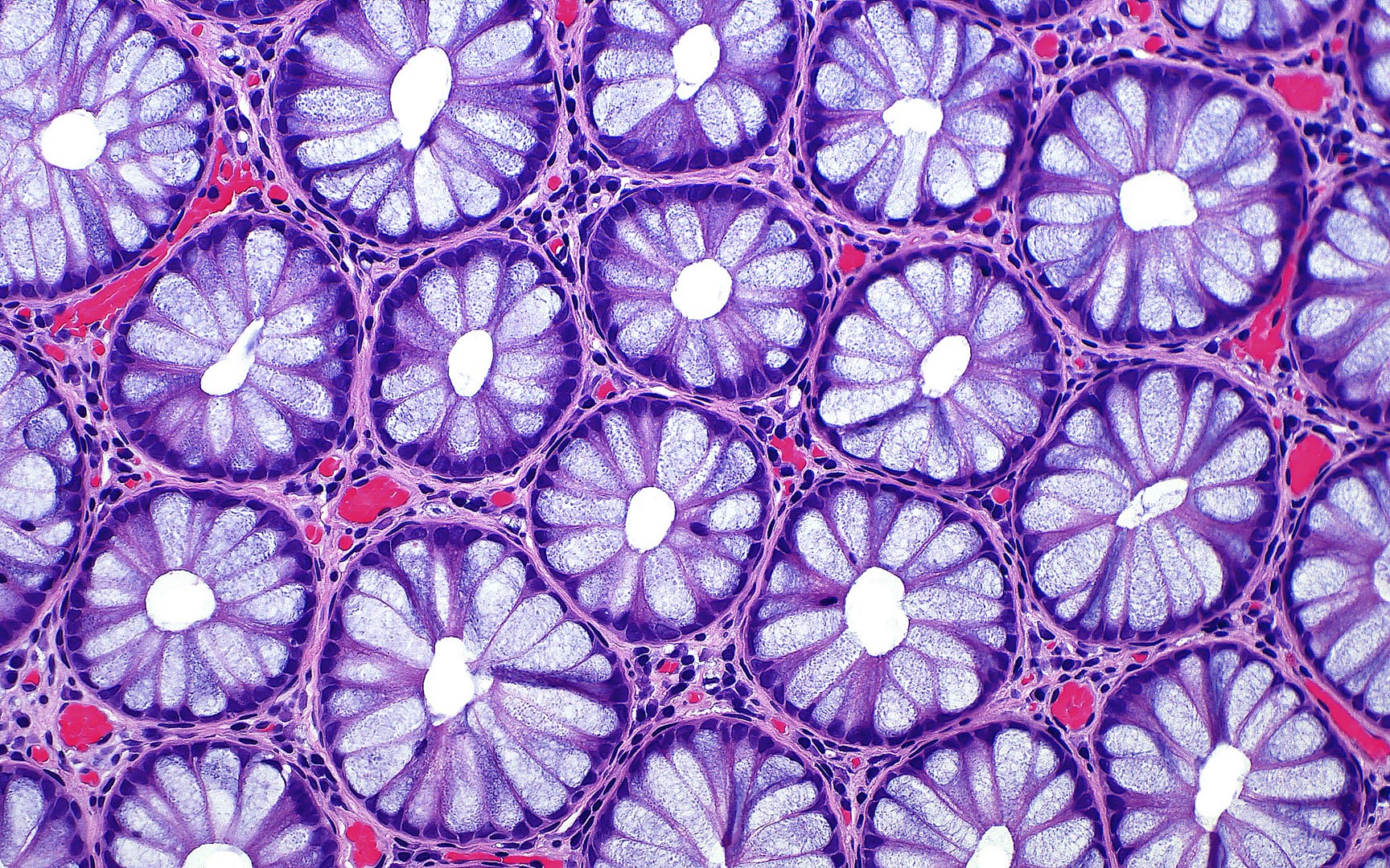
If any of these scientists get tired of studying the human colon, or red algae, or dinosaur bones (as above), there could be a bright future for them in textile design. But really, all the images reveal incredible worlds largely hidden from public view, brought to light by science.
“Each year, Nikon Small World receives an array of microscopic images that exhibit exemplary scientific technique and artistry. This year was no exception,” said Nikon spokesperson Eric Flem. “At the intersection of art and science, this year’s competition highlights stunning imagery from scientists, artists, and photomicrographers of all experience levels and backgrounds from across the globe.”
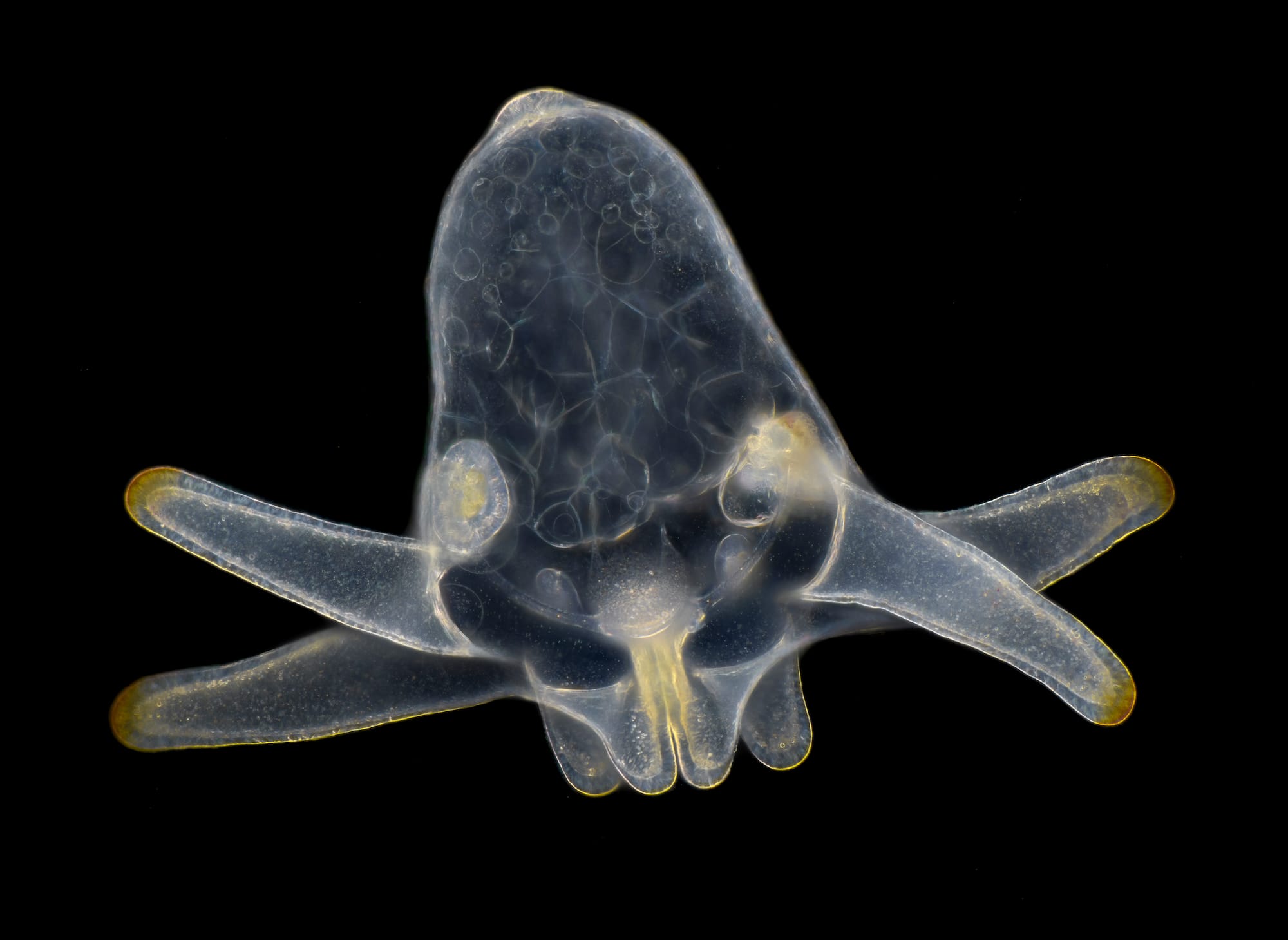
And as this adorable image of the larva of an anemone in marine plankton demonstrates, even the smallest of gestures can create a big picture.