The First Printed Illustration of a Modern Dissection
Published in the late 15th century, the Fasciculus Medicinae contains the earliest depiction of a modern dissection, a groundbreaking representation for anatomy.

In a series for the first day of each month, Hyperallergic is exploring some firsts in art, from the earliest known depictions of things to pioneers in the visual fields.
To comprehend the human body, it’s necessary for the physician to understand its inner-workings. Yet dissection has long been a complex and controversial task. In 18th-century Great Britain, posthumous dissection was a punishment for criminals, and grave robbing in the 19th century for anatomical dissection often preyed on non-white and indigent burial grounds. The portrayal of dissections in art has reflected these anxieties. For instance, in Rembrandt’s “The Anatomy Lesson of Dr. Nicolaes Tulp” (1632), each face is incredibly detailed, including that of the corpse, a thief named Aris Kindt who had been hanged. It was important for the artist to communicate that this was not just an anonymous deceased.
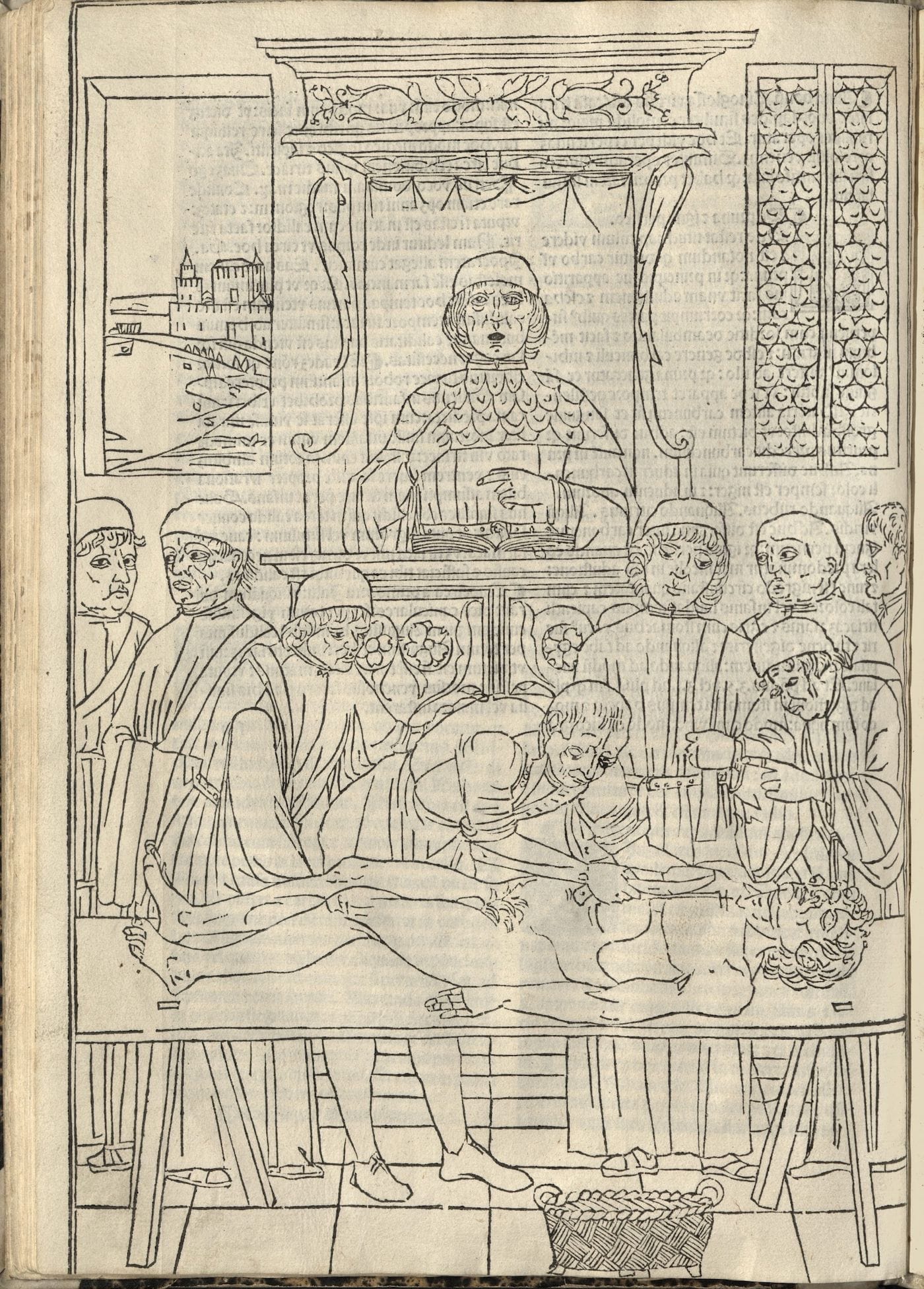
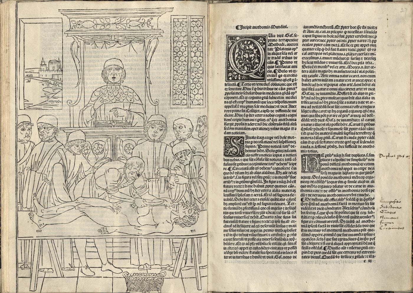
However, in the 15th century, the first printed depiction of a modern dissection portrayed the process with dignity. The woodcut in Fasciculus Medicinae, a compilation of medical treatises first published in 1491 in Venice by the brothers Gregorii and reprinted in 14 editions until 1522, captures the first cut on the corpse. Charles Singer described the scene in the 1925 second volume of Monumenta Medica:
In the fore part of the picture stands a trestle table on which a corpse is about to be dissected. On the ground in front of it is a basket to receive the viscera. The body is a nature-study, the bent legs, straight arms and risus sardonicus being all typical of the position assumed during rigor mortis. A physician acts as demonstrator; he bears a wand and points out the procedure. Before him stands the ostensor. This menial has his sleeves tucked up and in his right hand he bears the knife with which he is about to make the incision. Around the couple, thus actively engaged in the operation, stand six physicians in academic dress. Their finely drawn faces, which are evidently portraits, are among the triumphs of the illustrator of this volume.
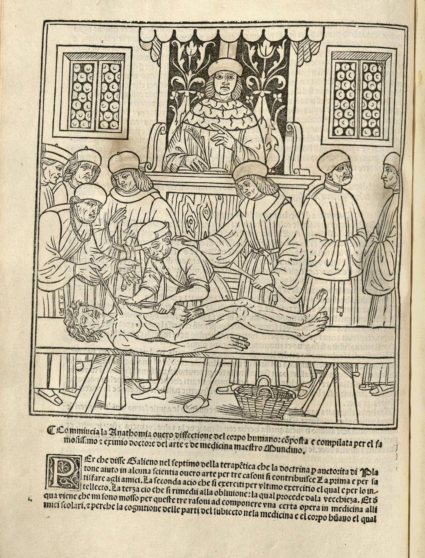
Anne Garner, curator of rare books and manuscripts at the New York Academy of Medicine (NYAM) Library, told Hyperallergic that the dissection illustration in Fasciculus Medicinae “makes sense because it accompanies a text by Mondino de Luzzi, ‘On Anatomy,’ about dissection. This section begins, ‘Here begins the anatomy, or dissection of the human body, composed and compiled by the most famous and esteemed doctor of the art, etc. of medicine, Maestro Mondino.'”
The NYAM Library has five editions of Fasciculus Medicinae, the earliest being from 1495, and is currently working on a digital exhibition called Facendo Il Libro/Making the Book that will explore its art and text. “Many other images of dissection appear in anatomical atlases from the 16th century on,” Garner explained. “The body on the table appears in earlier printed editions of the Mondino de Luzzi text, printed before the turn of the 16th century — the body is just not yet dissected in the earlier edition.”
Although tension over who was dissected existed in the 15th and 16th centuries, there was a major interest in accurately representing the anatomy of the human body (see Leonardo da Vinci’s incredible sketches of flayed cadavers). “Dissection images absolutely become common, and it doesn’t take long,” Garner stated. “Vesalius’s Fabrica — the milestone anatomical work of the 16th century — features Vesalius himself performing a dissection in the anatomical theater. In 1685, Bidloo’s anatomical atlas shows the dissected body with props: ropes, pins, dissection tools, tables, etcetera, even a fly on a cadaver, rendering the realities of the dissection room in 105 copperplate engravings.”
Fasciculus Medicinae is considered to contain the earliest realistic anatomical images in print, such as an illustration known as the “Zodiac Man” where different areas of the body are connected to zodiac signs. (Garner shared a scan of it from a 1522 edition on the NYAM blog.) It’s joined by a text that acts as a guide to when bloodletting should be done. Although your zodiac likely has little influence today on when you elect to get an operation or blood test, much in the 15th century manuscript echoes the current practice of medicine, including the quest for knowledge through the examination of the dead.


