"I Have Seen My Death": The First X-Ray Photograph
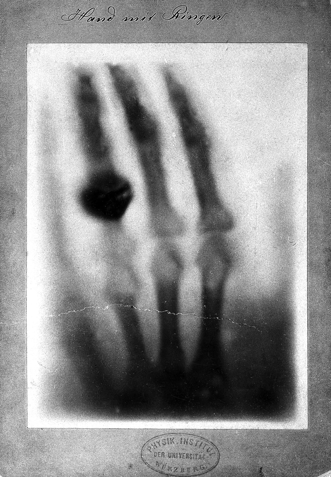
In December of 1895, Wilhelm Röntgen revealed the bones of his wife's hand in the first X-ray photograph.

In a series for the first day of each month, Hyperallergic is exploring some firsts in art, from the earliest known depictions of things to pioneers in the visual fields.
When Anna Bertha Ludwig saw the bones of her hand exposed beneath her skin, her wedding wing hovering above the skeletal knuckles, she reportedly exclaimed, “I have seen my death.” The memento mori visual, later immortalized on a photographic plate, is known as the first medical X-ray image. It was created on December 22, 1895 by her husband Wilhelm Röntgen, who had discovered this penetrating electromagnetic radiation that November.
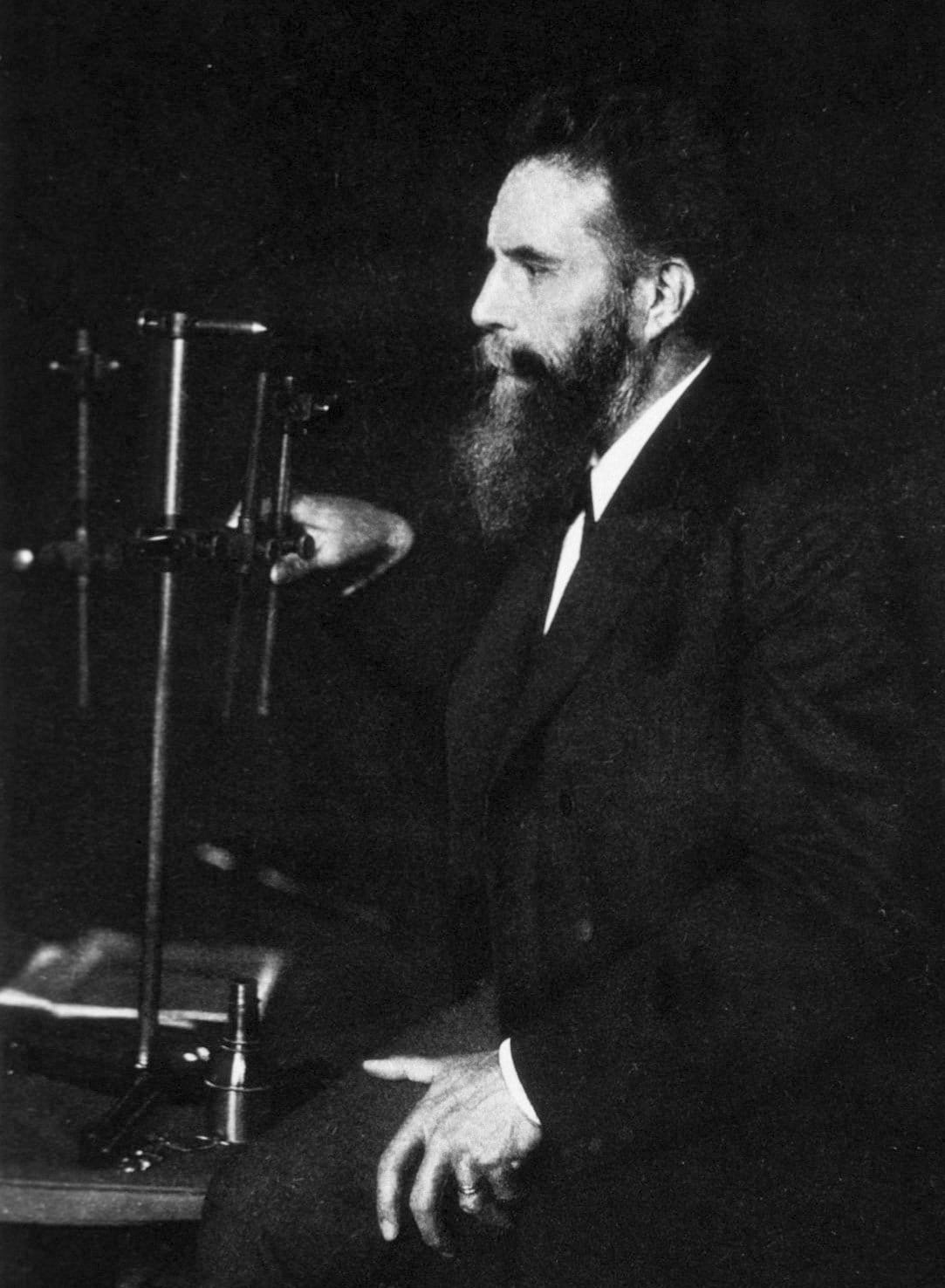
While Anna was haunted by the exposure of her inner self, that ability to see within the human body without incision radically improved the life-saving capabilities of medicine in the 20th century. In January 1896, Röntgen presented his “X-ray,” so named for its then-unknown properties that he’d found while experimenting with cathode rays, to the Würzburg Physico-Medical Society. There he created a plate of an attending anatomist’s hand to demonstrate its capabilities. While the proposed name “Röntgen’s rays” did not stick, the technique caught on quickly, and the physicist was awarded the Nobel Prize in 1901.
That visual of Anna’s skeleton hand was widely printed alongside the news of the discovery, such as a February 4, 1896 report in the New York Times (which followed a January 19 article that had dismissed this “alleged discovery of how to photograph the invisible”). It was an arresting image that conveyed immediately how the X-ray could pass through skin, but not metal, and reveal this anatomical world. The hand X-ray even became something of a radiograph, or “röntgenogram,” fad, and you can find images of the jewelry-laden bones of the Baroness Josephina Mollinary-Vranyczany from 1896, and Alexandra, Empress of Russia, from 1898.
Today, Anna’s hand appears on a commemorative coin, stamp, and even a mural on the Röntgens’ former Utrecht home. Of course, there were victims of overexposure in these early X-ray labs; for instance, Clarence Dally, X-ray assistant to Thomas Edison, died in 1904 at the age of 39 from metastatic skin cancer. Yet the X-ray had an incredible influence on 20th-century medical treatment, from skull fractures of World War I to the questionable use of the machines for shoe-fitting in the midcentury, and all trace back to that startling image of a woman’s hand.


